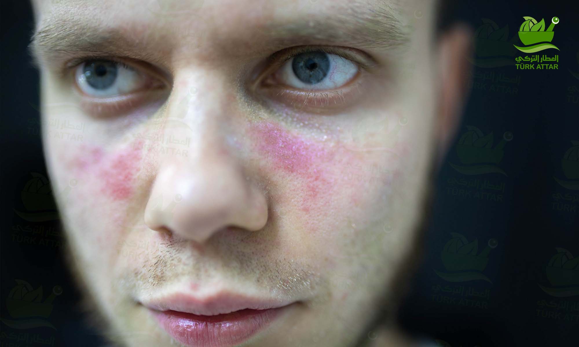
الذئبة الحمامية الجهازية هي مرض مناعي ذاتي جهازي مزمن لسبب غير معروف، يتميز بالعديد من النتائج المتعلقة بالتهاب العديد من الأنسجة والأعضاء مثل الجلد والمفاصل والكلى والتأمور وغشاء الجنب والعديد من تشوهات الجهاز المناعي (المناعية).
الذئبة الحمامية الجهازية
تم وصف المرض لأول مرة على أنه اضطراب جلدي مزمن من قبل طبيب الجلد الفرنسي بيت في عام 1833. مصطلح الذئبة يعني "الذئب" في اللاتينية ويشير إلى خاصية تدمير الأنسجة في الآفة. لاحظ Kaposi في عام 1872 أن المرض كان جهازي. ظاهرة "خلية الذئبة" هي نتيجة مهمة في تشخيص المرض وصفها Hargraves في عام 1948. لاحقًا ألقى اكتشاف الأجسام المضادة الذاتية المضادة للنواة (ANA) بواسطة Frio في عام 1957 الضوء على سمة أمراض المناعة الذاتية للذئبة الحمامية الجهازية (SLE).
كيف يحدث المرض؟
في مرضى الذئبة الحمراء يكون الجهاز المناعي غير طبيعي من جميع النواحي لذلك من غير المعروف ما هي الشذوذ الضروري في تكوين مرض الذئبة الحمراء. يُعتقد أن العوامل البيئية تلعب دورًا في بدء واستمرار مرض الذئبة الحمراء في الأفراد المهيئين وراثيًا. تم ملاحظة حدوث زيادة في الإصابة بمرض الذئبة الحمراء في بعض العائلات في الشعوب السوداء والشرق الأقصى والأمريكيين الأصليين. إذا كان أحد أفراد الأسرة مصابًا بمرض الذئبة الحمراء فإن خطر الإصابة بمرض الذئبة الحمراء في التوائم المتماثلة يزداد بنسبة 30٪ تقريبًا وبنسبة 5٪ للأقارب الآخرين من الدرجة الأولى.
تسود فكرة أن العوامل البيئية تلعب دورًا من خلال إثارة خلل في التنظيم المناعي لدى الأفراد الذين لديهم استعداد وراثي. من بين هذه العوامل، يمكن حساب الفيروسات والأشعة فوق البنفسجية والأدوية.
بعض الأدوية مثل بروكاييناميد، هيدرالازين، فينيتوين، آيزونيازيد، تسبب إنتاج ANA ويمكن رؤية مشابهة سريريًا لـ SLE تُعرف هذه الحالة بمرض الذئبة أو متلازمة شبيهة الذئبة.
تسبب معظم العوامل المعدية تحفيز المناعة وإنتاج السيتوكين وقد يتسبب في حدوث الذئبة لدى الأفراد الذين لديهم استعداد وراثي. قد يكون النقص الخلقي للبروتينات التكميلية موجودًا في مرض الذئبة الحمامية، يعد نقص C2 أكثر شيوعًا من غيره.
قد تلعب أوجه القصور التكميلية دورًا في ظهور المرض من خلال خلق الحساسية للإصابة بالعدوى بالإضافة إلى ذلك لا ينبغي أن يكون مفاجئًا أن الزيادة في نشاط الخلية البائية (الخلية التي تصنع الأجسام المضادة) ضرورياً أيضًا في تكوين مرض الذئبة الحمراء.
تتمثل الآلية المعروفة لتطور المرض بوساطة الأجسام المضادة في ترسب معقدات الأجسام المضادة للمستضد في الأنسجة، تم إثبات الترسبات بشكل خاص في الأوعية وكبيبات الكلى. تشكل الأجسام المضادة الذاتية المطورة ضد البروتينات داخل الخلايا والأحماض النووية مجمعات مناعية منتشرة عن طريق الارتباط بالمستضدات المنبعثة من الخلايا الميتة. معرفتنا بالمستضد محدودة، لكن غالبًا ما يكون نوع الجسم المضاد IgG. يؤدي ترسب المعقدات المناعية في الأنسجة يسبب التنشيط والاستجابة الالتهابية. من خلال مكونات C3a و C5a المتممة، يتم تنشيط الخلايا الالتهابية وإطلاق المواد الالتهابية، ويؤدي تنشيط خلايا التخثر إلى تكوين جلطة صغيرة، وإنتاج مستقلبات الأكسجين التفاعلية، وإطلاق إنزيمات التحلل المائي والسيتوكينات تسبب تلفاً مباشراً للأنسجة. يؤدي الوجود الدائم للمجمعات المناعية إلى تلف الأنسجة المزمن. سريريًا ينتج عنه التهاب الأوعية الدموية والتامور وغشاء الجنب والآفات الجلدية والتهاب الكلى. تشكيل ندب في الأعضاء المصابة بالالتهاب ولوحظ فقدان الوظيفة في الأعضاء المصابة بالالتهاب.
الجنس الأنثوي هو أيضًا عامل خطر مهم لتطور مرض الذئبة الحمامية الجهازية. تم الكشف الآن عن التشوهات في استقلاب الإستروجين (الهرمون الأنثوي) والأندروجين (هرمون الذكورة) التي تظهر في المرضى الذين يعانون من مرض الذئبة الحمامية الجهازية ونماذج الفئران المصابة بمرض الذئبة. على وجه الخصوص تم الكشف الآن عن الدور المهم لهرمون الاستروجين في التسبب في المرض.
الإصابة:
مرض الذئبة الحمامية ليس مرضا نادرا ففي السنوات الأخيرة أدى تطوير الاختبارات المناعية الحساسة على التعرف على الأشكال الخفيفة من المرض بواسطة الأجسام المضادة للنواة والأجسام المضادة للحمض النووي والمكملات إلى زيادة معدل الإصابة. تم الإبلاغ عن أن معدل انتشار المرض هو 15-50 لكل مائة ألف. المناطق الجغرافية المختلفة لديها نسبة منخفضة أو عالية من مخاطر الإصابة. المرض أكثر شيوعًا 3-4 مرات في العرق الأسود من العرق الأبيض.
على الرغم من أن مرض الذئبة الحمامية الجهازية يمكن أن يحدث في أي عمر إلا أنه أكثر شيوعًا بين سن 13- 40. 90٪ من المرضى هم من النساء في سن الإنجاب. نسبة الإناث إلى الذكور 9/1. يظهر مرض الذئبة أيضًا عند الأطفال وكبار السن وهو أكثر شيوعًا بين الفتيات ثلاث مرات منه عند الأولاد.
النتائج السريرية:
نادرًا ما يُلاحظ ظهور مرض الذئبة الحمامية الجهازية النموذجي في بعض المرضى ففي كثير من الأحيان، يعاني المرضى في البداية من عرض أو عرضين، مثل التعب والتهاب المفاصل الروماتويدي ثم قد تتطور أعراض أخرى لـ SLE. تختلف الأعضاء المصابة بالمرضى وتختلف شدة المرض باختلاف نظام العضو المعني. يتميز مرض الذئبة الحمامية بفترات تفاقم وهدوء. عند التشخيص يعاني معظم المرضى من أعراض أساسية مثل التعب والحمى وفقدان الوزن. دعونا الآن نفحص كل هذه النتائج واحدة تلو الأخرى.
ما يقرب من 90٪ من مرضى الذئبة الحمامية يكون العرض الأول هو التهاب المفاصل (التهاب المفاصل) أو ألم المفاصل (آلام المفاصل) خاصة المتماثل مع انتفاخ عرضي للأنسجة الرخوة. الأقل شيوعًا هو التهاب المفاصل (التهاب أكثر من مفصل واحد). عادة قد تتأثر المفاصل والمعصمين والمرفقين والكاحلين. تم ملاحظة تصلب الصباح في 50٪ من المرضى. قد تكون النتائج الالتهابية في المفصل مؤقتة أو مزمنة. عادة ما تكون التغيرات المدمرة (النموذجية لمرض التهاب المفاصل الروماتويدي) غائبة في التهاب المفاصل SLE. من المحتمل أن تكون التشوهات ناتجة عن إصابة المفاصل المزمنة.
تم ملاحظة آلام العضلات عند 1/3 من المرضى في بداية المرض وبعض المرضى لديهم حساسية في العضلات. قد يكون هناك أيضًا ضعف في العضلات وانخفاض في الأنسجة العضلية. يُلاحظ مرض العضلات بسبب العلاج بأدوية الكورتيزون أو الملاريا.
تشوهات الجلد والشعر والأغشية المخاطية هي ثاني أكثر الأعراض شيوعًا لمرض الذئبة الحمامية الجهازية (85٪ من المرضى). يمكن رؤية العديد من الأنواع المختلفة من المظاهر الجلدية في مرض الذئبة. يمكن أن يحدث طفح جلدي أحمر اللون على شكل فراشة (طفح جلدي) والذي يغطي الخدين وجسر الأنف ولا يظهر في الأخاديد بين الأنف والشفتين دون التعرض لأشعة الشمس ومع ذلك يمكن أن تزداد مع ضوء الشمس. الطفح الجلدي الثاني الأكثر شيوعًا في مرضى الذئبة هو طفح جلدي مرتفع يمكن أن يكون في أي مكان على الجسم. غالبًا ما يسبق تفاقم الآفات الجلدية التفاقم الجهازي للمرض. بالإضافة إلى الآفات المذكورة أعلاه يمكن أيضًا رؤية أعراض جلدية أخرى مثل الشرى والفقاعة (الحويصلات المليئة بالمصل) وشبكي حي (مظهر يشبه الخريطة) والتهاب السبلة الشحمية (التهاب الأنسجة الدهنية تحت الجلد) وتساقط الشعر. تحدث تقرحات الغشاء المخاطي للفم والتي غالبًا ما تكون غير مؤلمة في الحنك الرخو والصلب. ظاهرة رينود (تبييض اليدين أو القدمين في البرد والاحمرار بعد الكدمات) يمكن أن تكون شديدة بما يكفي لتسبب الغرغرينا.
تم العثور على حساسية للضوء (حساسية الضوء) في 50-60٪ من المرضى بالإضافة إلى زيادة الآفات الجلدية المصاحبة لأشعة الشمس، يمكن أيضًا ملاحظة زيادة في النتائج الجهازية.
ما يقرب من 50 ٪ من المرضى يعانون من تورط كلوي مهم سريريًا. الفشل الكلوي هو سبب مهم للوفاة في مرضى الذئبة الحمامية.
تظهر النتائج العينية في حوالي 20٪ من المرضى.
قد تحدث إصابة الرئة أو القلب أو الصفاق في مرض الذئبة الحمامية. تم العثور على إصابة الغشاء الرئوي في 30-60٪ من المرضى. يعاني المريض من آلام جانبية تزداد مع التنفس والسعال.
يحدث التهاب التامور بشكل أقل من التهاب ذات الجنب (20-30٪). على الرغم من أن التهاب التامور لا يتم اعتباره سريريًا، إلا أن ECO يمكنه اكتشاف السوائل في تجويف الغشاء.
على الرغم من أن التهاب الصفاق (التهاب الغشاء البريتوني) ليس شائعًا فقد وجد أن 60٪ في تشريح الجثث. في المرضى الذين يعانون من ظهور مفاجئ للغثيان والقيء وآلام البطن المنتشرة، يمكن النظر في احتمال الإصابة بالتهاب الصفاق.
في مرض الذئبة الحمامية الجهازية تشارك جميع طبقات القلب بالتساوي في المرض. التهاب الشغاف (التهاب البطانة الداخلية للقلب) هو المظهر القلبي النموذجي لهذا المرض. على الرغم من أنها صامتة في الغالب فقد تم اكتشافها في 30 ٪ من دراسات التشريح. يمكن أيضًا رؤية مرض صمام القلب في مرض الذئبة. كإحدى اكتشافات الأوعية الدموية، يصاب 10٪ من المرضى بتخثر داخل الأوعية الدموية معظمهم في الساقين.
كما أن أعراض الجهاز العصبي مختلفة تمامًا لدى هؤلاء المرضى. بالإضافة إلى الأعراض مثل الذهان والاكتئاب، يمكن رؤية نوبات الصرع والنزيف الدماغي والشلل المؤقت عند المرضى. قد يرتبط الاكتئاب والذهان من النتائج النفسية أيضًا باستخدام الكورتيزون في هذه الحالة من الضروري التوقف عن الدواء.
تم الكشف عن نتائج الجهاز الهضمي في 50٪ من المرضى. يعد فقدان الشهية والغثيان والقيء من أكثر الأمراض شيوعًا. قد تكون هذه النتائج بسبب التهاب الصفاق أو أمراض الأوعية الدموية في الأمعاء أو العلاجات الدوائية. تظهر إصابة الجهاز الهضمي كعلامات للمريء، التهاب الأوعية المغذية للأمعاء، أمراض الأمعاء الالتهابية، التهاب البنكرياس أو أمراض الكبد.
تم ملاحظة حجم طحال خفيف أو معتدل في 20٪ من المرضى. خلال الفترات النشطة سريريًا للمرض يحدث تضخم منتشر في العقدة الليمفاوية في نصف المرضى، هذه النتيجة أكثر شيوعًا عند الأطفال. تتغير أيضًا اضطرابات خلايا الدم مع نشاط المرض. النتيجة الأكثر شيوعًا هي فقر الدم. لوحظ تدمير كبير لخلايا الدم في 10٪ من المرضى. بصرف النظر عن هذا يمكن أيضًا رؤية التشوهات والانخفاضات في خلايا الدم الأخرى.
ANA ليس خاصًا بـ SLE. فالنتائج الإيجابية تشير إلى مرض الذئبة الحمامية. ANA 95-98٪ إيجابية في مرض الذئبة يمكن اعتبار المستويات العالية من مضادات dsDNA من عائلة ANA محددة لهذا المرض. توجد في 75٪ من المرضى.
المستويات التكميلية (C3 و C4) تكون منخفضة في المرضى النشطين.
في مرض الكلى النشط توجد البروتينات والتراكيب والخلايا الحبيبية والقوالب في البول.
كيف يتم التشخيص؟
يجب الاشتباه في مرض الذئبة الحمامية لدى الأشخاص المصابين بمرض متعدد الأجهزة مع آلام المفاصل.
